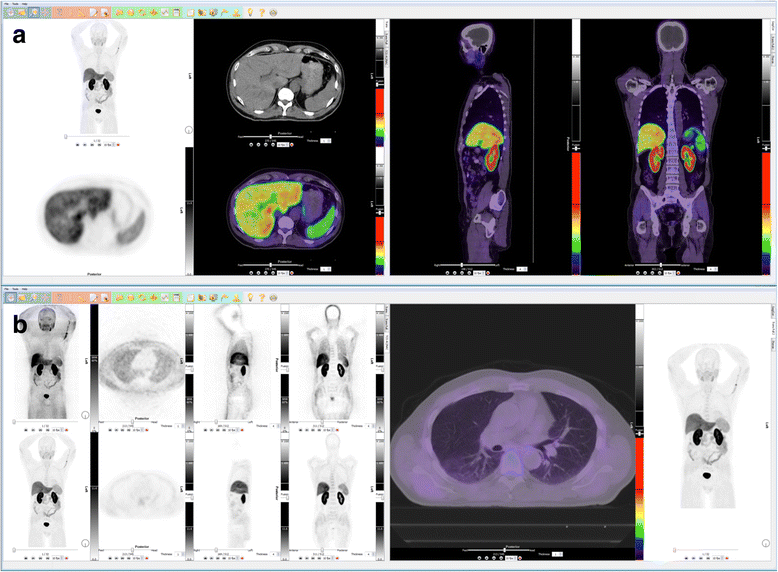
Fig. 1

Dual-screen whole-body FCH-PET/CT image display template (Hybrid Viewer, Hermes Medical Solutions, Stockholm, Sweden). The primary tab (a) includes AC PET MIP plus CT, PET and fused PET/CT transaxial views on the left screen, and fused sagittal and coronal views on the right screen. The secondary tab (b) includes MIP and orthogonal views of NAC and AC PET on the left, and a large transaxial PET/CT view on the right. A rainbow color LUT (Siemens ECAT Rainbow) is used for the fused images, which are displayed with a linear, 50–50% blend. AC PET imaged intensity is normalized by adjusting the upper limit of the color scale so that the liver is nearly, but not, saturated (mostly yellow to orange). The same SUV upper threshold is used to adjust the inverse gray scale intensity of the PET-only images, as well as the dynamic PET images. This is a case of a negative FCH-PET/CT in a patient with biochemical relapse after radical prostatectomy, where physiological FCH distribution is seen