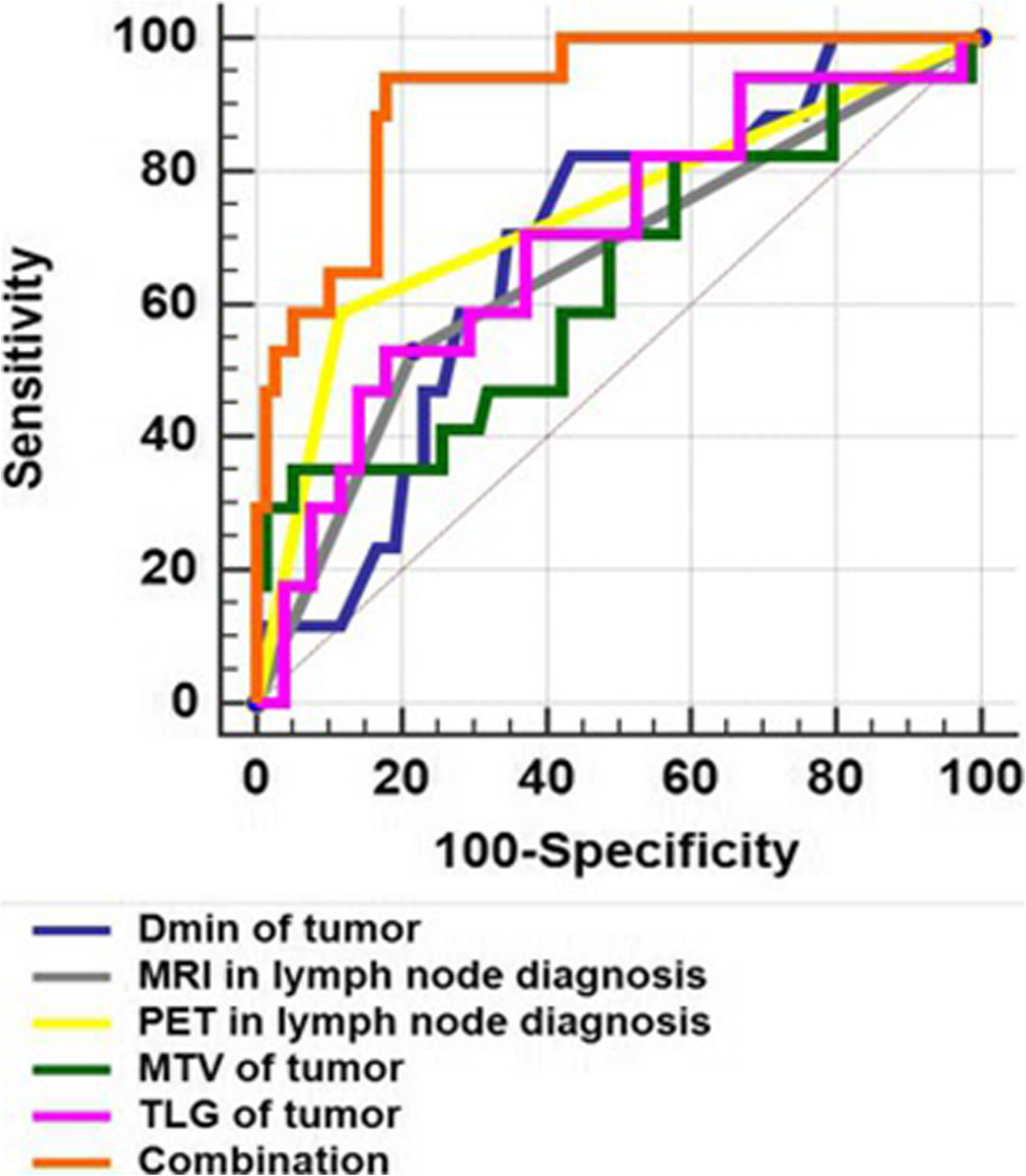
Fig. 5

For all patients, ROC analysis shows that SUVmax (AUC 0.654, 95% confidence interval (CI) 0.549–0.749, p = 0.023), SUVmean (AUC 0.646, 95% CI 0.541–0.741, p = 0.030), TLG (AUC 0.692, 95% CI 0.588–0.782, p = 0.009), Dmin (AUC 0.681, 95% CI 0.577–0.773, p = 0.007), MRI in lymph node diagnosis (AUC 0.656, 95% CI 0.551–0.750, sensitivity 58.94, specificity 78.21, p = 0.020) and PET in lymph node diagnosis (AUC 0.736, 95% CI 0.636–0.822, sensitivity 58.82, specificity 88.46, p < 0.001) had a positive effect on predicting metastatic lymph nodes confirmed by postoperative pathology. The area under the ROC curve for the combination of TLG, Dmin and PET/MRI in lymph node diagnosis (AUC 0.913, 95% CI 0.837–0.961, sensitivity 94.12, specificity 82.05, p < 0.001) was higher than any individual parameter (both, p < 0.05). CI: confidence interval